

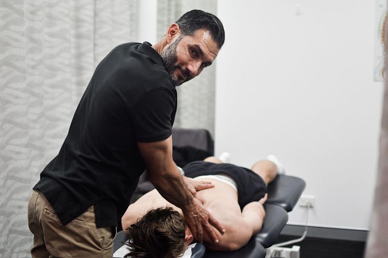
Health Blog
In the Media
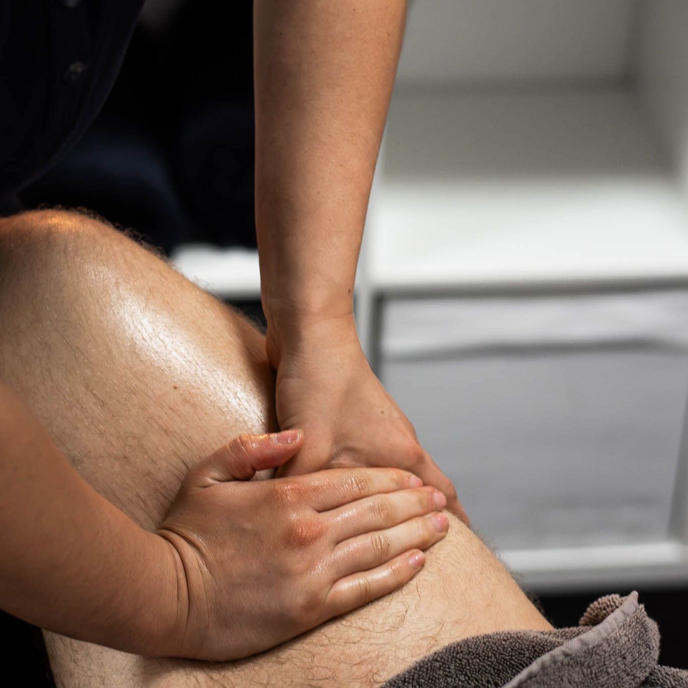
Elastoplast Taping & Movement Series
Episode 11: Gluteal Muscle Strengthening and Control
Elastoplast Taping & Movement Series
Episode 4: Ankle Taping and Rehabilitation
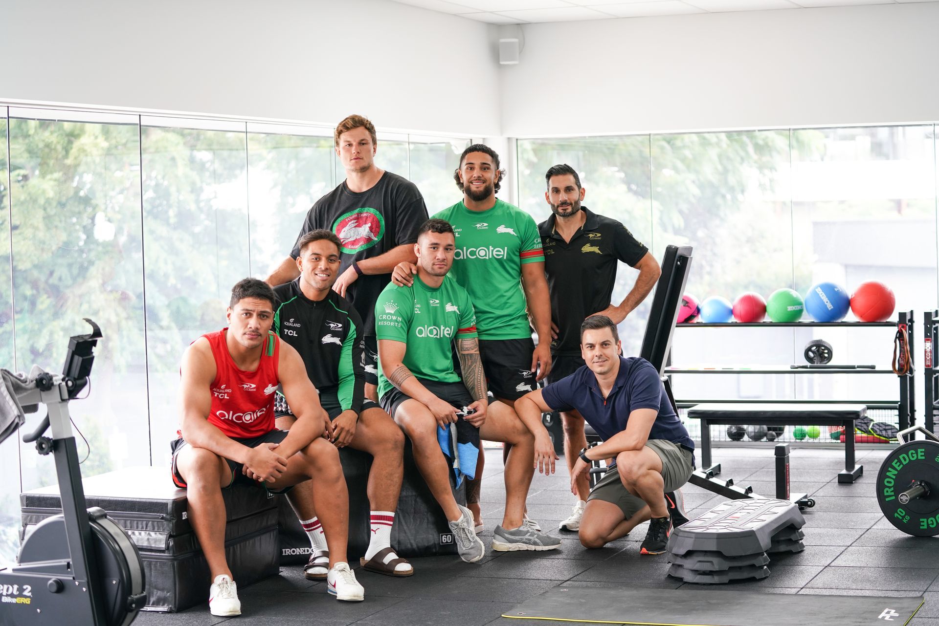
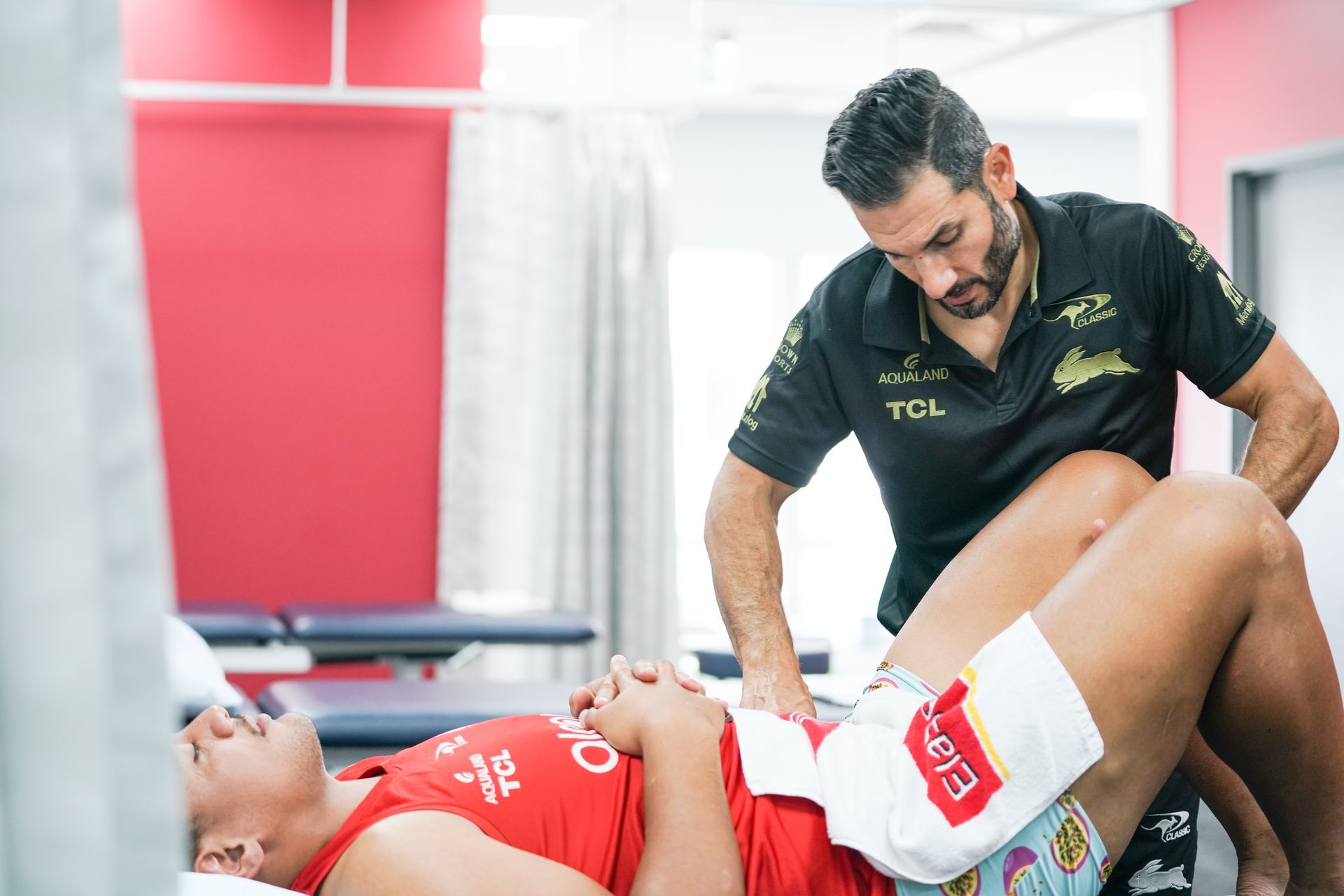
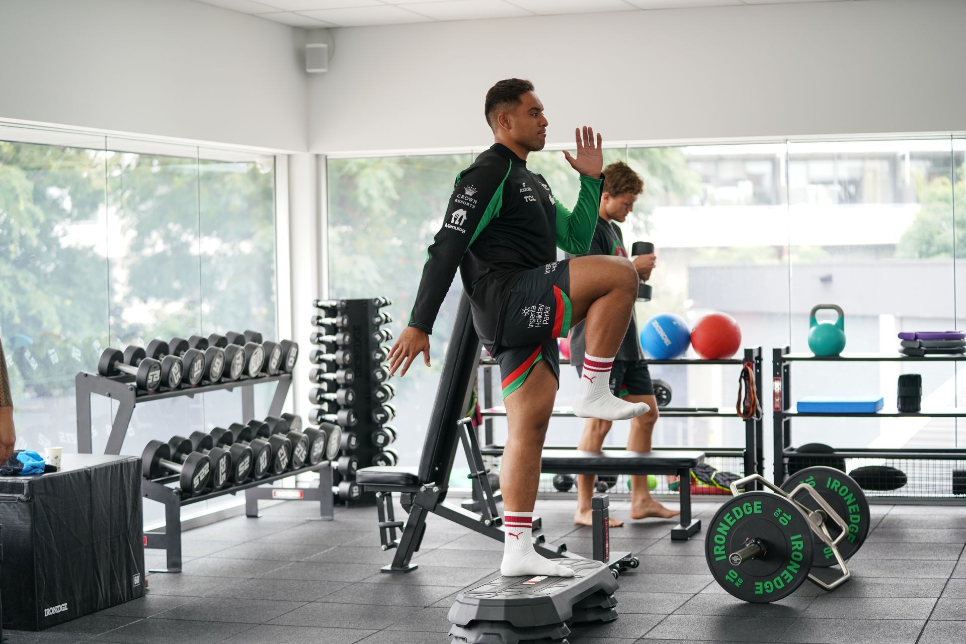
South Sydney Herald
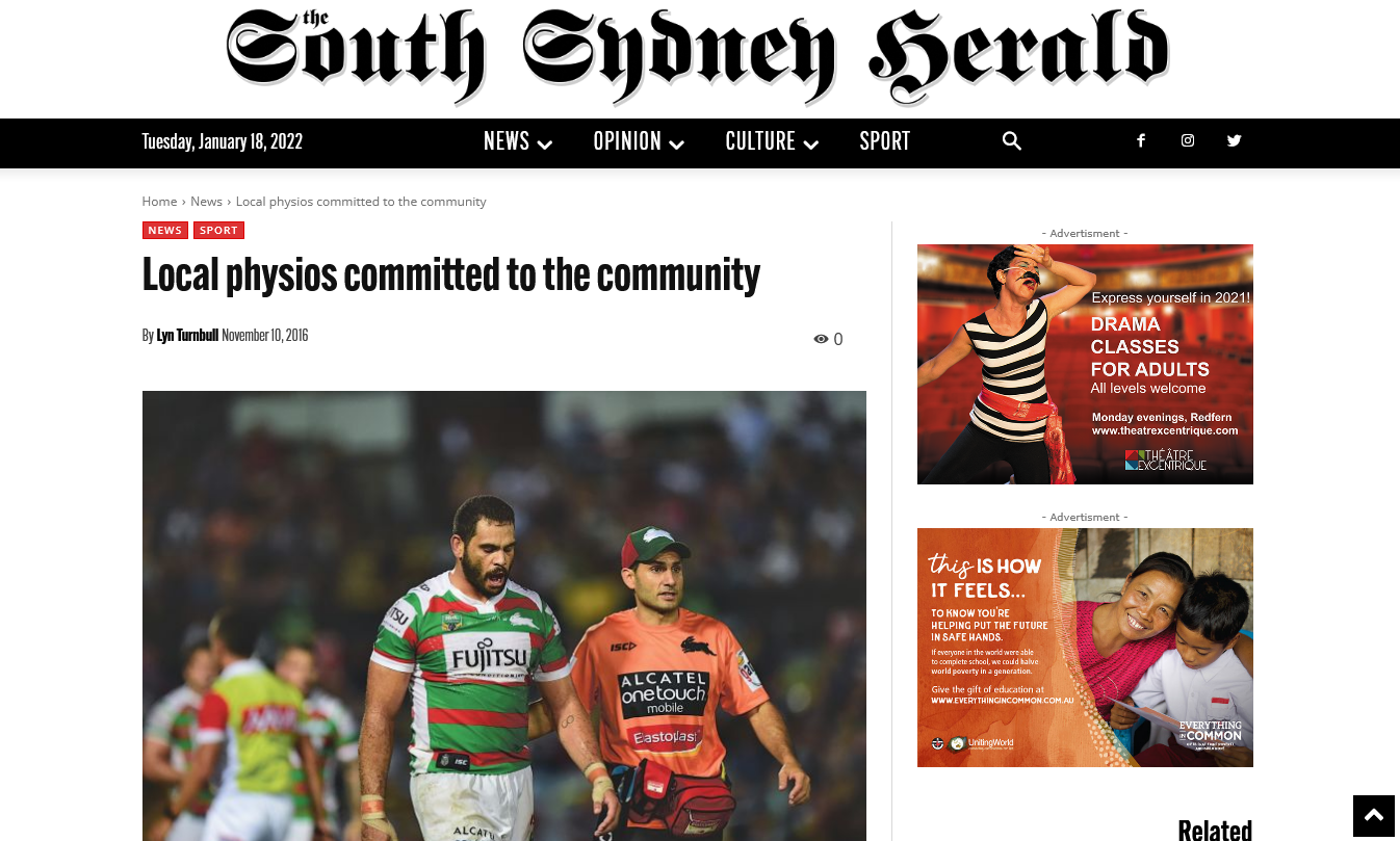
Local Physios Committed to the Community
Our latest health blogs

Special Considerations for Hamstring Injuries Hamstring injuries are a common injury among athletes, particularly those involved in sports that require sudden changes of direction, explosive acceleration, and high-speed running. Rehabilitation after a hamstring injury can be a challenging process, and it is essential to understand the special considerations involved in the rehabilitation process to ensure optimal outcomes. The latest evidence has shown hamstring injuries require assessment and treatment of multiple areas of the body such as lumbopelvic control, posterior chain strength and stability and hamstring strength in a fatigued state. Due to the complex nature of hamstring injuries and their relationship with the aforementioned areas, they have a high recurrence rate. In this blog, we will discuss the special considerations for hamstring rehabilitation. The Importance of Early Intervention One of the most critical special considerations for hamstring rehabilitation is the importance of early intervention. Delaying rehabilitation can prolong the recovery time and increase the risk of re-injury. Therefore, it is essential to initiate rehabilitation as soon as possible after the injury. Gradual Progression Another important consideration for hamstring rehabilitation is the need for gradual progression. It is crucial to progress rehabilitation exercises slowly and steadily, focusing on regaining strength, flexibility, and mobility while avoiding overloading the hamstring muscles. Eccentric Exercises Eccentric exercises are particularly effective for hamstring rehabilitation. These exercises involve lengthening the muscle while it is under tension, which helps to promote muscle fibre remodelling and healing. Eccentric exercises can also help to improve muscle strength and reduce the likelihood of recurrence. Sports-Specific Rehabilitation Rehabilitation exercises should be tailored to the specific requirements of the athlete's sport. For example, athletes involved in sports that require high-speed running may benefit from incorporating high-speed running drills into their rehabilitation program. This approach can help to improve neuromuscular control, endurance, and power. How Can High-Speed Running Help with Hamstring Rehabilitation? The use of high-speed running as a rehabilitation strategy for hamstring injuries is based on the principle of progressive loading. High-speed running involves significant eccentric loading of the hamstring muscles, which can help to stimulate muscle fibre remodelling and improve muscle strength and endurance. Evidence for High-Speed Running for Hamstring Rehabilitation Several studies have explored the use of high-speed running as a rehabilitation strategy for hamstring injuries, with promising results. A systematic review published in the British Journal of Sports Medicine evaluated the effectiveness of high-speed running in the rehabilitation of hamstring injuries. The review analysed seven studies with a total of 301 participants. The review found that high-speed running was effective in improving hamstring strength, running performance, and reducing the risk of re-injury. The review also found that high-speed running did not increase pain or re-injury risk compared to traditional rehabilitation programs. Another study published in the Journal of Orthopaedic and Sports Physical Therapy evaluated the effectiveness of high-speed running in the rehabilitation of elite soccer players with hamstring injuries. The study found that high-speed running was effective in reducing the time to return to play, improving hamstring strength, and reducing the risk of re-injury. The Role of Manual Therapy Manual therapy can also be beneficial for hamstring rehabilitation. Soft tissue mobilisation techniques, such as soft tissue release, can help to improve blood flow, reduce muscle tension, and promote tissue healing. Joint mobilisation techniques around the lumbar spine and pelvis can also help to improve joint mobility, reducing the risk of further injury. This is integral as we work on ensuring the lumbopelvic region is working efficiently to allow the hamstrings to rehabilitate to their full potential. Psychological Considerations Recovering from a hamstring injury can be a frustrating and challenging process, and psychological consider a tions should also be taken into account during rehabilitation. It is common for people to experience the highs and lows of rehab & may benefit from working with a sports psychologist to help them cope with the emotional and psychological challenges of rehabilitation and stay motivated throughout the process. This is also integral for return to sport considerations as it can be a daunting experience after injury. Concerns of re-injury or not being able to perform at their pre-injury level is often felt and thus it is important for people to feel confident and mentally prepared to return to sport following a hamstring injury. Conclusion Hamstring rehabilitation requires special considerations to ensure optimal outcomes. Early intervention, gradual progression, eccentric exercises, sports-specific rehabilitation, manual therapy, and psychological considerations are all essential elements of an effective rehabilitation program. It is essential to work with a physiotherapist to develop a personalised rehabilitation program tailored to the athletes specific needs and goals.
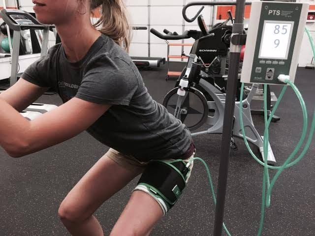
Application and benefits during rehab. Blood flow restriction (BFR) training involves restricting the blood flow to working muscles during exercise, resulting in hypoxia in the muscle tissue. It can be applied to both resistance and aerobic exercise; however, the purpose of this blog is to have a look at the potential benefits of using BFR during resistance-based exercise in rehab. BFR resistance training has been shown to help increase muscle size and strength in young, old and load compromised populations (including post ACL replacement surgery and people suffering from OA). The greatest benefits of BFR training have been shown to occur during low-load resistance exercise, with some studies showing that muscle hypertrophy and strength adaptions using BFR are significantly greater than those achieved with low load resistance exercise alone. Benefits of BFR during rehab After sustaining an injury or having surgery, people will often struggle tolerating heavy loads through the affected area, which can cause an increase in pain during activity, reduce function and impact rehab and recovery timeframes. During rehab for any type of injury, it is important to load and strengthen the muscles around the injured or painful structure, however pain (and injury) can often prevent a person from being able to perform the required exercises (or tolerate the required load during exercise) to actually achieve this increase in strength, which can often lead to slow progression during rehab. This is where BFR training can be beneficial. BFR training can be used to get the most out of low-load resistance training for load compromised people who can’t tolerate heavy-load training during the early stages of rehab. Applying BFR training to rehab As mentioned above, BFR training can be a useful tool when trying to maximise the impact of low-load exercise during rehab. This can often be beneficial when pain/injury is impacting the ability to appropriately load the injured area in order to elicit a change in symptoms. A good example of this presentation in the clinic is during rehab following a patella dislocation or subluxation. The sensitivity and reduced capacity to load the patellofemoral joint after this type of injury can make it hard to perform quadriceps strengthening exercises with adequate load to elicit a meaningful change in strength, which can lead to the development of compensatory strategies and slow progression during rehab. BFR can be used to help overcome this hurdle. Have a look at our Instagram here to see an example of a rehab session using BFR for a client who suffered a patella dislocation. There are many other types of pain/injury that could benefit from the use of BFR at certain stages of rehab, such as managing OA pain and when recovering from ACL replacement surgery. Overall, BFR can be a useful tool to use during certain stages of rehab; but just like all aspects of rehab there is no ‘one size fits all’ approach, so get in contact in you would like to discuss if BFR could be useful for you. Reference for information Hughes L, Paton B, Rosenblatt B, Gissane C, Patterson SD. Blood flow restriction training in clinical musculoskeletal rehabilitation: a systematic review and meta-analysis. Br J Sports Med. 2017 Jul;51(13):1003-1011. doi: 10.1136/bjsports-2016-097071. Epub 2017 Mar 4. PMID: 28259850.

The ‘High Ankle Sprain’ - Syndesmosis Injuries Syndesmosis injuries, also known as high ankle sprains, are a type of injury that occurs in the ankle joint. Unlike traditional ankle sprains, which affect the ligaments on the outside of the ankle, syndesmosis injuries involve the ligaments that connect the tibia and fibula bones in the lower leg. These ligaments are known as the syndesmotic ligaments, and they help to stabilise the ankle joint during movement. Symptoms of syndesmosis injuries can include pain, swelling, and tenderness on the front of the ankle. The pain is often located above the ankle joint and can be severe, especially during weight-bearing activities. In more severe cases, the individual may experience difficulty bearing weight on the affected leg and may have limited mobility in the ankle joint. Syndesmosis injuries are often caused by a twisting or rolling motion of the ankle, which can occur during sports or other physical activities. They can also be caused by a fall or other trauma to the ankle joint. Individuals who participate in sports that involve jumping, cutting, or pivoting movements are at higher risk for syndesmosis injuries. Diagnosis of syndesmosis injuries usually involves a physical examination of the ankle joint, as well as imaging tests such as X-rays or MRI scans. The following is an example of a sports rehab program for an individual recovering from a syndesmosis injury: Phase 1: Acute Phase (1-2 weeks) Rest, ice, compression, and elevation to manage pain and swelling Non-weight bearing with the use of crutches Gentle range of motion exercises such as ankle circles and ankle pumps Isometric exercises for ankle strengthening Phase 2: Sub-Acute Phase (2-4 weeks) Weight-bearing as tolerated Active range of motion exercises such as calf loading and calf raises Strengthening exercises such as calf raises, single leg balance exercises and posterior chain loading Manual therapy techniques such as soft tissue massage and joint mobilisation Sport-specific training Phase 3: Functional Phase (4-6 weeks) Plyometric exercises to improve power and agility Sport-specific drills to improve coordination and balance Proprioception and balance training Advanced strengthening exercises such as squats, lunges, and lateral movements Return to sport or activity with gradual progression and careful monitoring It is important to note that every individual's rehab program will be different and will depend on the severity of their injury, as well as their overall health and fitness level. It is recommended to work closely with a qualified physiotherapist to ensure a safe and effective rehab program that is tailored to your specific needs and goals. A systematic review published in the British Journal of Sports Medicine in 2019 found that early functional rehabilitation, which includes range of motion, strength, and proprioception exercises, can lead to better outcomes and faster return to sport for individuals with syndesmosis injuries compared to immobilization or surgery alone. Another study published in the Journal of Orthopaedic Surgery and Research in 2019 found that a structured rehabilitation program that included progressive weight-bearing, range of motion, and strength exercises resulted in significant improvements in pain, function, and ankle range of motion for individuals with syndesmosis injuries. In summary, syndesmosis injuries are a type of ankle injury that can be caused by a variety of factors. Prompt diagnosis and treatment are important for a successful recovery, and rehabilitation programs can help individuals return to their normal activities safely and effectively. By taking steps to prevent ankle injuries in general, individuals can reduce their risk of experiencing a syndesmosis injury and maintain good overall ankle health.

ACL Prehab Injuring your ACL can be overwhelming, often you will be deliberating whether or not to get surgery while coming to terms with the long rehab process ahead. This blog will shed a bit of light on something that everyone should be doing after sustaining an ACL injury; Prehab. What is ACL Prehab? ACL Prehab is what is done between tearing your ACL and having surgery/performing rehab. It is the term used for the exercises and training that needs to be performed after sustaining an ACL injury and should be performed even if you are considering not having surgery. What does ACL Prehab involve? Prehab involves a block of supervised exercise sessions with a focus on lower body strengthening (particularly the quadriceps and hamstrings), knee mobility and stability/motor control. The timeframes and specific types of exercise will vary between different people, but the goal of prehab remains the same; maximise the chance of returning to your pre-injury level of activity and staying there. Why is Prehab so important? Performing a 5-week block of prehab training has been shown to increase the chance of returning to your pre-injury level of sport following an ACL replacement, it has also been shown to significantly improve knee function after ACL injury, regardless of whether you decide to have surgery or not (Eitzen et al, 2010). Prehab also provides a great opportunity to perform some strength and hop testing, which will be used later in rehab. This testing will provide a good comparison for leg strength and function prior to returning to sport, doing this has been shown to help reduce the risk of reinjury by up to 75% (Capin et al, 2019). The take home message. A short block of Prehab has been shown to have a great impact on knee function after ACL injury and will increase the chances of having a good recovery and returning to your chosen sport or activity, regardless of whether or not you require surgery. Everyone who suffers an ACL injury should perform some amount of Prehab; if you have recently injured your ACL and would like to have a chat about Prehab, then get in touch.
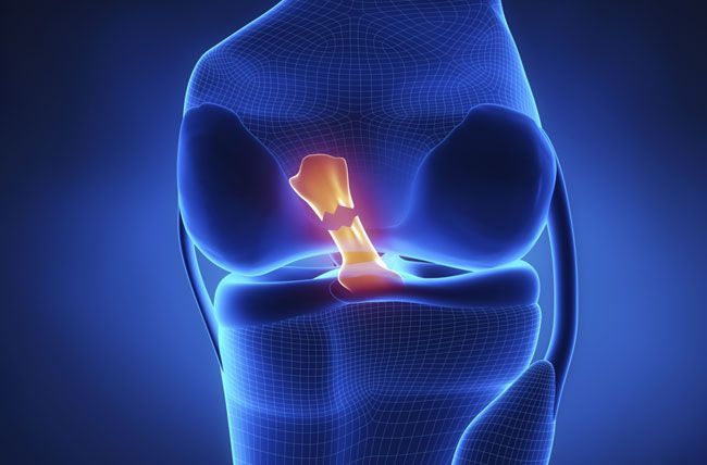
Torn ACL The Anterior Cruciate Ligament (ACL) is the most important stabilising structure within the knee joint, most commonly injured through either a contact (eg being tackle) or non-contact (change of direction) mechanism. The classic symptoms are often reported to be hearing/feeling a pop in the knee joint, immediate swelling, and pain as well as feelings of instability. Typically, hands on clinical assessments are very good at diagnosing an ACL rupture, however, imaging such as MRI is often used to assess whether there are any secondary injuries to other structures within the knee which may impact management. We are so used to hearing about ACL injuries being season ending events reported within most sports media outlets, with many athletes going on to have around a 9–12-month rehabilitation before returning to sport. It was widely accepted at one time that there are only 2 options for rehabilitation when someone has torn their ACL, either to have surgical reconstruction of the ACL ligament, using either a hamstring or patella tendon graft, or conservatively managing without an intact ligament through strengthening the muscles around the knee to provide stability. However, there have been recent studies which have shown some spontaneous healing of ACLs on MRI imaging in patients who have undertaken conservative rehabilitation (Costa-Paz et al. 2012, Filbay et al. 2022) So, it begs the question, surgical or conservative management? Well, what if there was a 3rd option? The Cross Bracing Protocol Exciting new findings were presented by Dr Tom Cross, a local Sydney Sports Physician, during a lecture held by the Australian Physiotherapy Association exploring the utilisation of a novel bracing protocol to promote natural healing of the ACL. The protocol is described as 4 weeks in a Range of Motion brace locked at 90 degrees of knee bend, with the next 8 weeks the brace slowly being let out to full knee extension. The reason for this is that by placing the knee in that position, it allows for the greatest chance for the two torn ends of the ACL to be aligned as closely as possible and hopefully lead to healing. Dr Cross’ preliminary work has shown that by following this protocol, 82 out of 84 patients demonstrated a healed ACL at 6 months post injury. Whilst there are many factors that go into which patients are offered the protocol and which have the greatest chance of healing, this case series demonstrates that there is an ability for the ACL to be able to heal on its own. This would allow for patients to be able to avoid possible risks of surgery, such as infection, whilst also having an intact ACL which would arguably allow them to return to sport in a quicker time frame. If you currently have an ACL injury or any knee injury which requires rehabilitation, you can book online via the website or chat to our friendly staff on 9194 1800 to book a detailed initial assessment and management plan to get you back to doing the things you love! References: Costa-Paz, Ayerza, M. A., Tanoira, I., Astoul, J., & Muscolo, D. L. (2011). Spontaneous Healing in Complete ACL Ruptures: A Clinical and MRI Study. Clinical Orthopaedics and Related Research, 470(4), 979–985. https://doi.org/10.1007/s11999-011-1933-8 Filbay, Roemer, F., Lohmander, S., Turkiewicz, A., Roos, E. M., Frobell, R., & Englund, M. (2022). 32 Spontaneous healing of the ruptured anterior cruciate ligament: observations from the KANON trial. BMJ Open Sport & Exercise Medicine, 8(Suppl 1), A3–A3. https://doi.org/10.1136/bmjsem-2022-sportskongres.
Alexandria Physiotherapy & Sports Injury If you're looking for expert physiotherapy and top-tier injury management near Rosebery, Alexandria Physio is your trusted choice! As Rosebery physio experts, our team of elite physiotherapists has been helping a diverse range of individuals from all over Sydney, including the nearby areas of Zetland, Mascot, Alexandria, and Green Square, achieve their health and fitness goals for years. At Alexandria Physio, we pride ourselves on creating a welcoming, supportive, and inclusive environment. We understand and respect the diverse backgrounds, values, and cultures of our clients, ensuring that everyone feels comfortable and heard during their physiotherapy journey. Our compassionate team is dedicated to providing personalised care that aligns with your unique needs and lifestyle. Our clinic, located just a short drive from Rosebery, is renowned for providing exceptional sports injury treatment, rehabilitation, athletic screening, and recovery & wellness services. Whether you’re recovering from a sports-related injury or looking for preventative care to avoid future injuries, we’re here to help you get back on your feet stronger than ever. Why Choose Alexandria Physio—Rosebery Physio Experts for Injury Recovery? Our clinic’s approach blends advanced techniques, hands-on care, and personalised programs, making us the go-to experts for sports injuries, musculoskeletal pain, and post-op rehabilitation. From muscle strains and tendonitis to more complex issues like neck and back pain, we use targeted treatments that restore mobility and prevent long-term injury, making us Rosebery’s best physios for pain relief and recovery. Convenient Location Just Minutes from Rosebery Situated on Fountain Street in Alexandria, we’re easily accessible for those in Rosebery and surrounding suburbs like Zetland, Mascot, and Green Square. Our location is just a few minutes’ drive from key areas like Gardeners Road, The Cannery precinct, and Eastlakes Shopping Centre, making it convenient for students, professionals, and families looking for physio experts near Rosebery. Comprehensive Physiotherapy Services by Rosebery Physio Specialists At Alexandria Physio, we offer a wide range of services designed to treat your specific needs. Whether you're dealing with a recent injury or managing a long-term condition, our team of physiotherapists is equipped to help you achieve optimal health: Sports Injuries: Whether it’s a sudden injury or an overuse issue, our experts will develop a detailed and data-driven sports-specific program to restore strength and mobility. Neck and Back Pain: Our therapists are experienced in treating common neck and back issues that hinder daily activities and quality of life. Post-Op Rehabilitation: We offer structured rehab programs post-surgery to ensure smooth and effective recovery, no matter the procedure. Dry Needling: For relief from chronic pain, our dry needling techniques target trigger points in muscles to restore normal function. We also offer specialised services such as ergonomic assessments and onsite physiotherapy for businesses, as well as injury prevention and performance enhancement programs via our partner company, InTouch Physiotherapy. Book an Appointment with Leading Rosebery Physiotherapists Looking for the best physiotherapy and sports injury treatment in Rosebery? Book your appointment today with leading Rosebery physio experts and take the first step toward recovery. FAQs about Physiotherapy near Rosebery What conditions do your physiotherapists treat? We specialise in treating sports injuries, post-surgical rehabilitation, chronic pain, and musculoskeletal conditions. How do I book an appointment at Alexandria Physio? You can book online or call us at 02 9194 1800. Do you offer physiotherapy for workplace injuries? Yes, we provide ergonomic assessments, injury prevention programs, CTP and WorkCover-approved treatment plans. No matter where you're coming from, our dedicated team is here to support your journey to better health. You don’t have to travel far to receive the best physiotherapy and sports injury care – book your appointment online today or contact us to learn more about how we can help you recover and thrive.
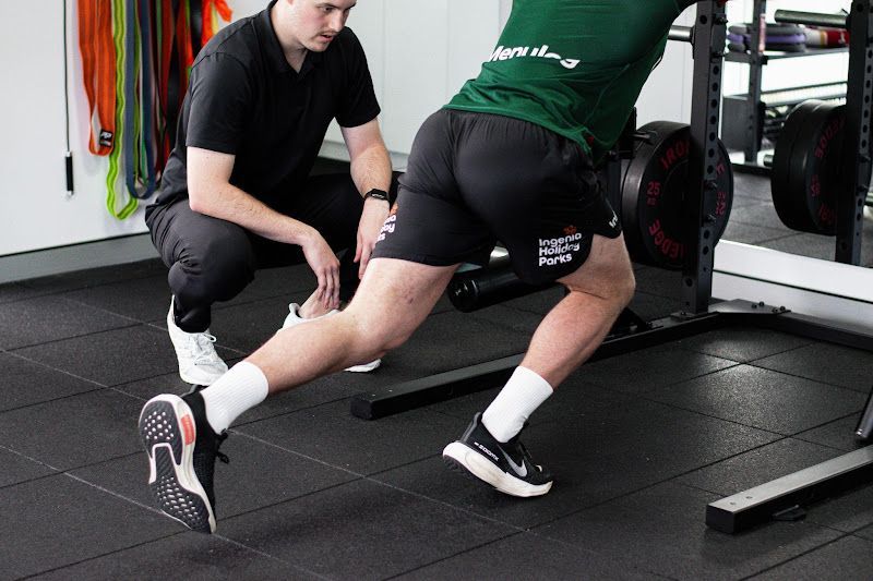
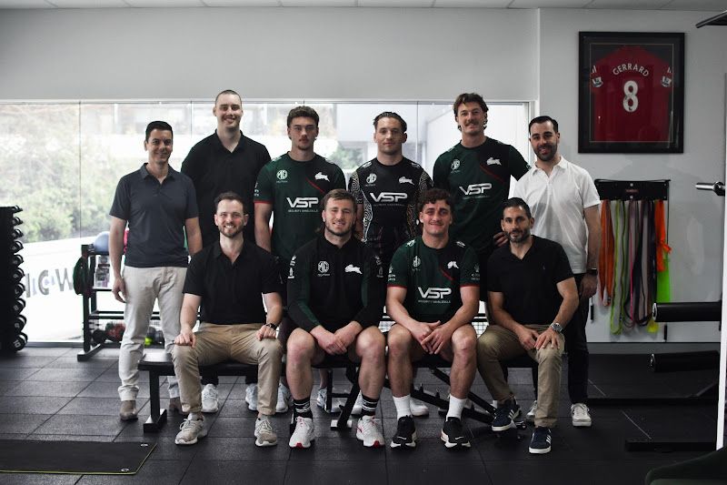
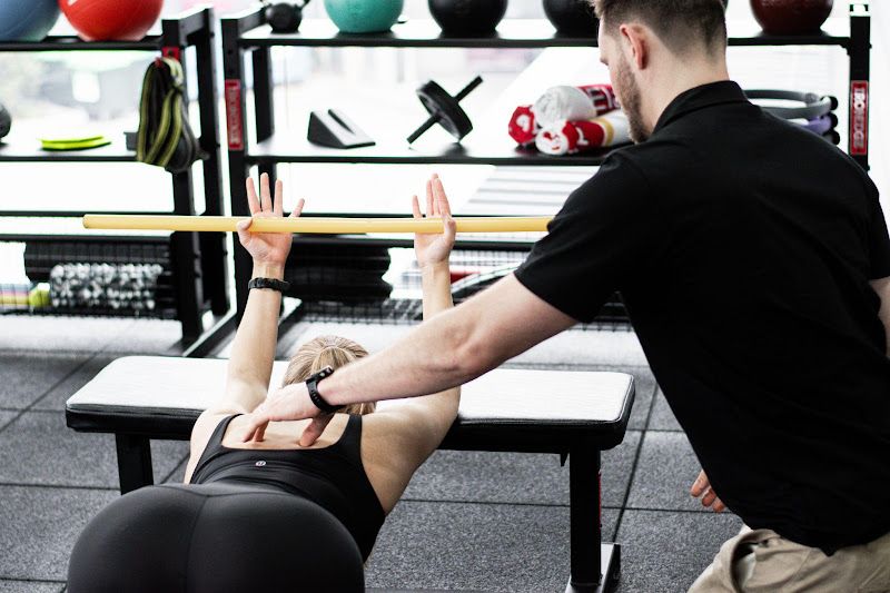
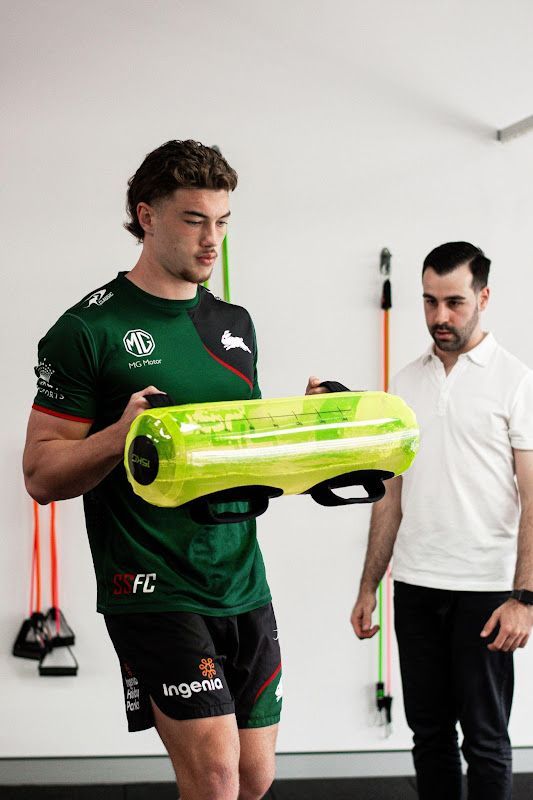
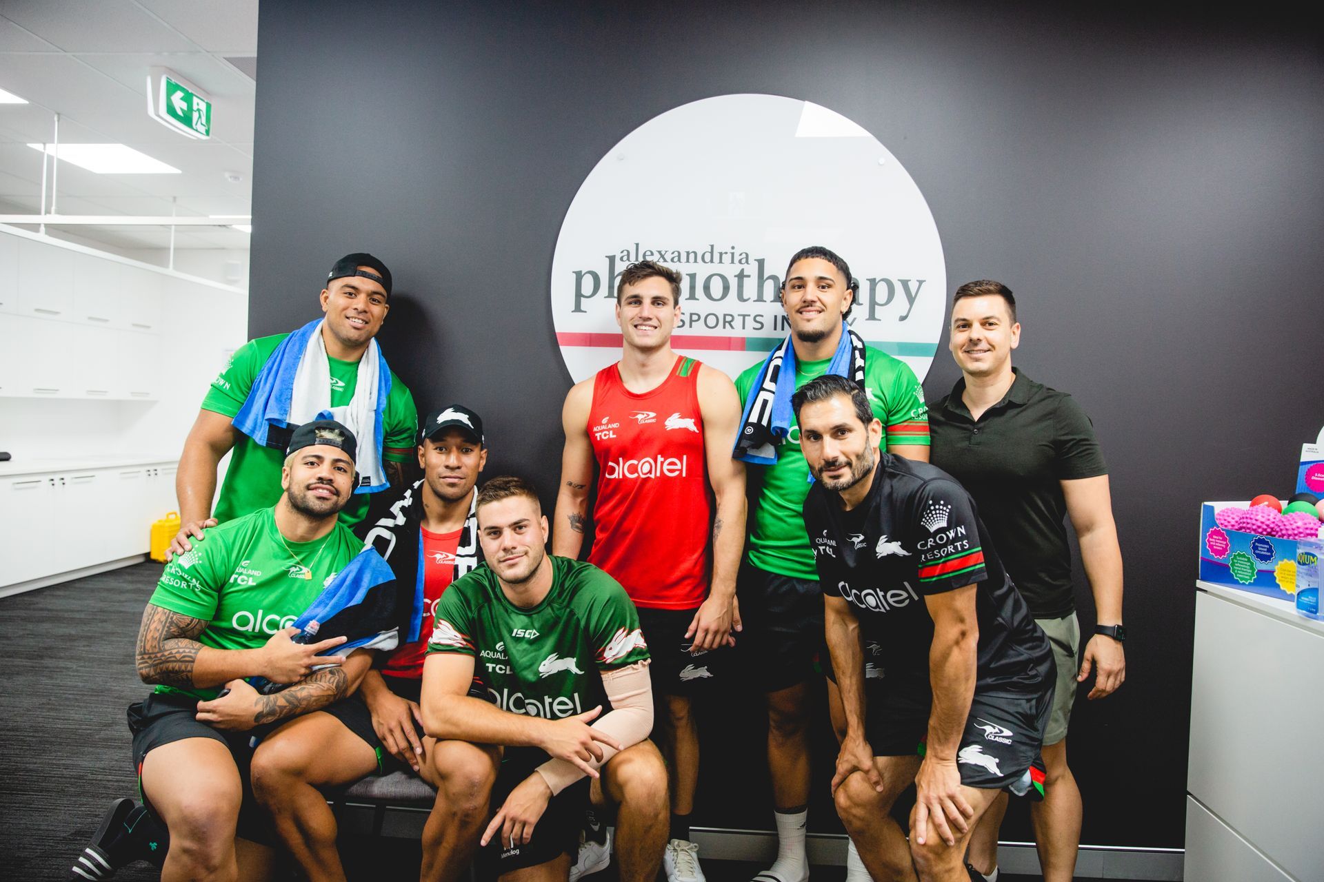
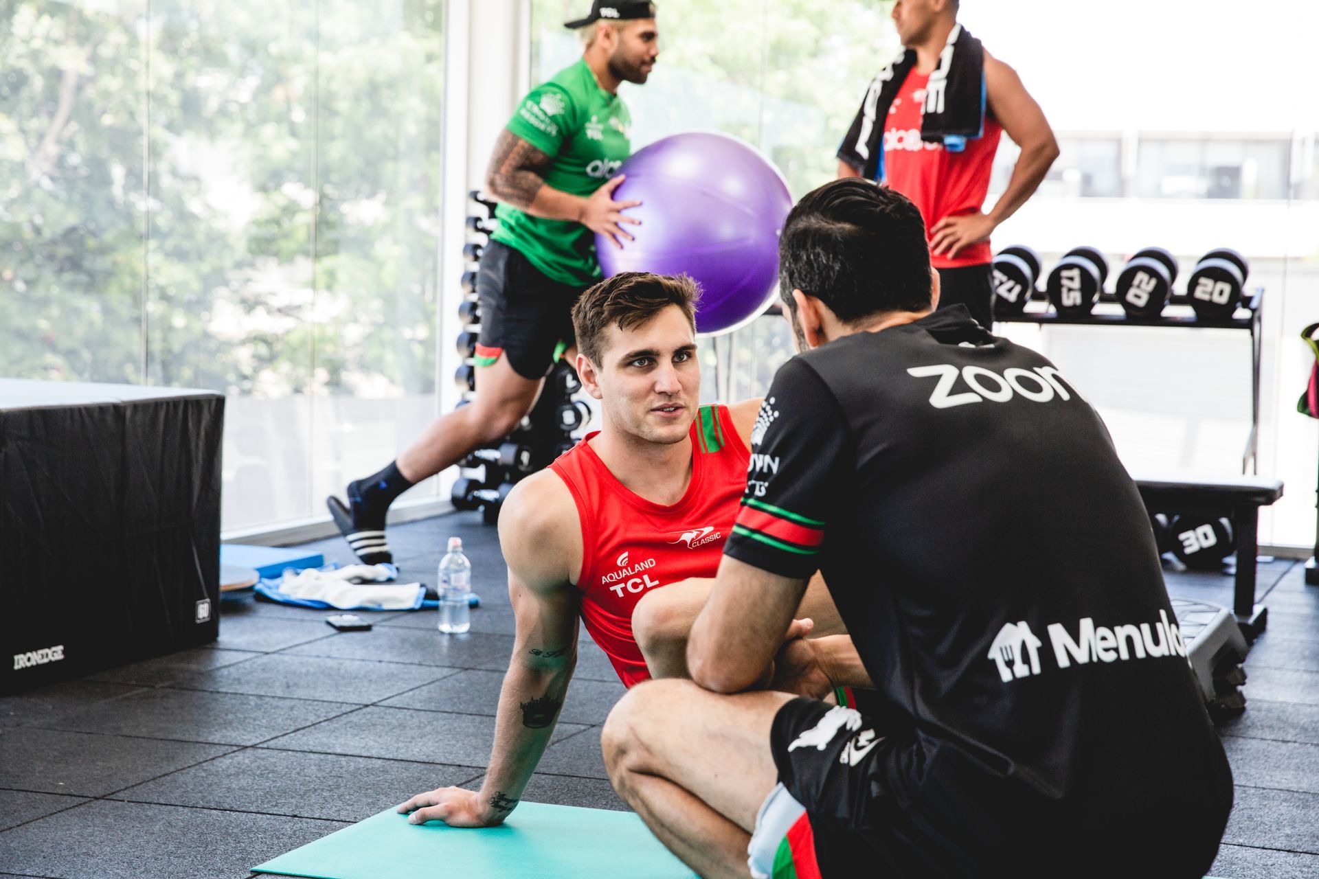
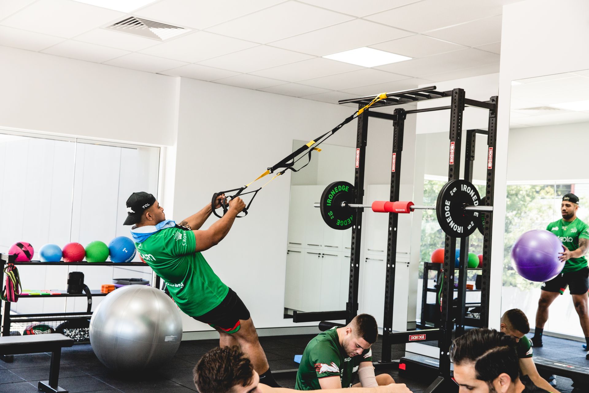
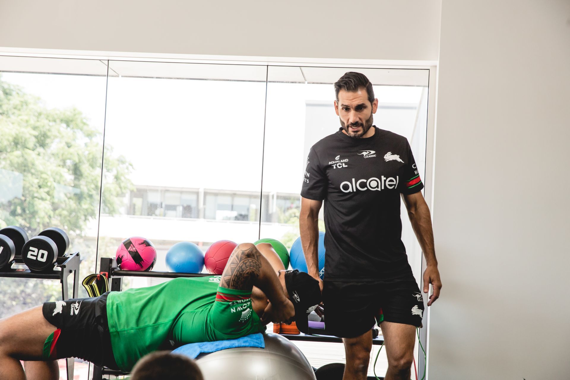
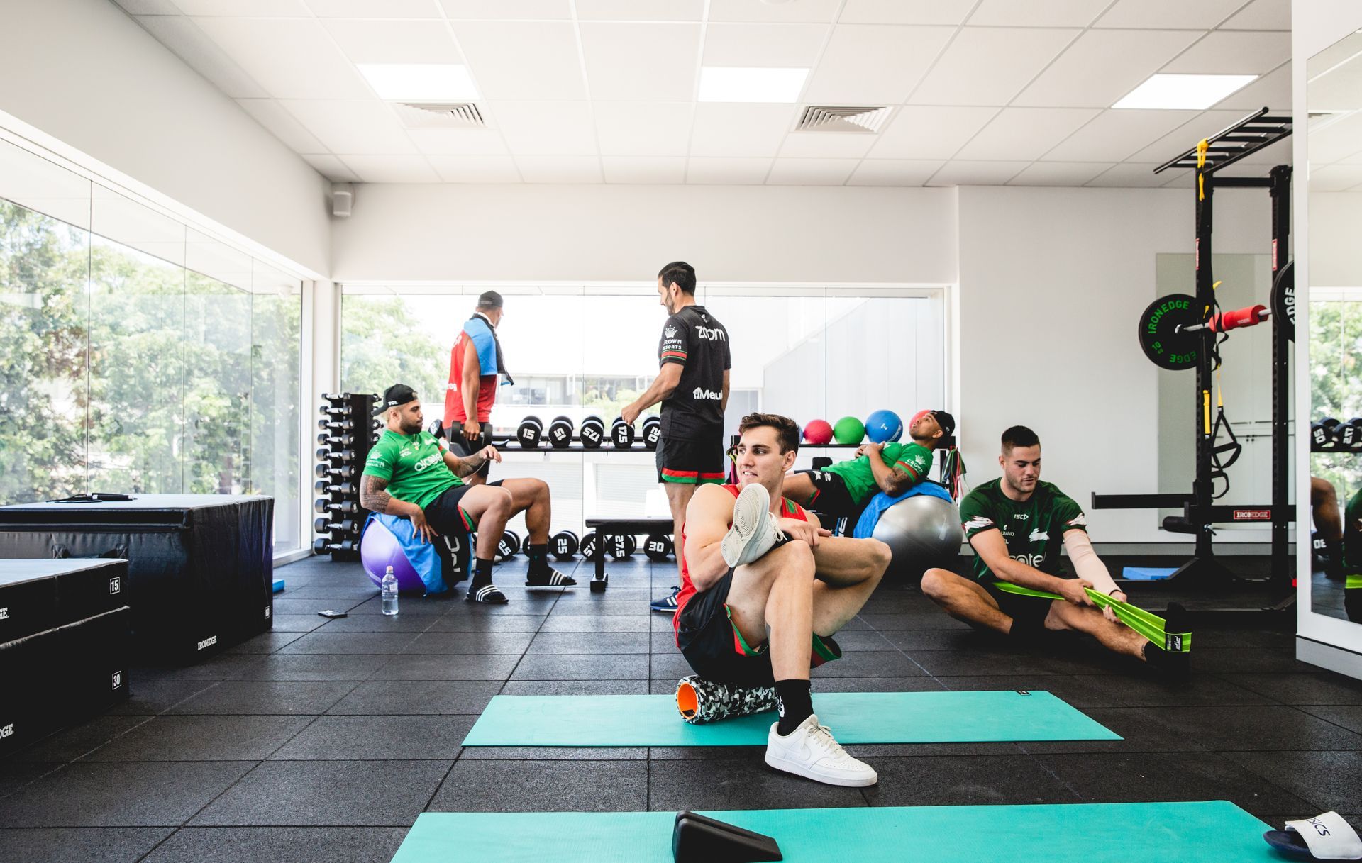
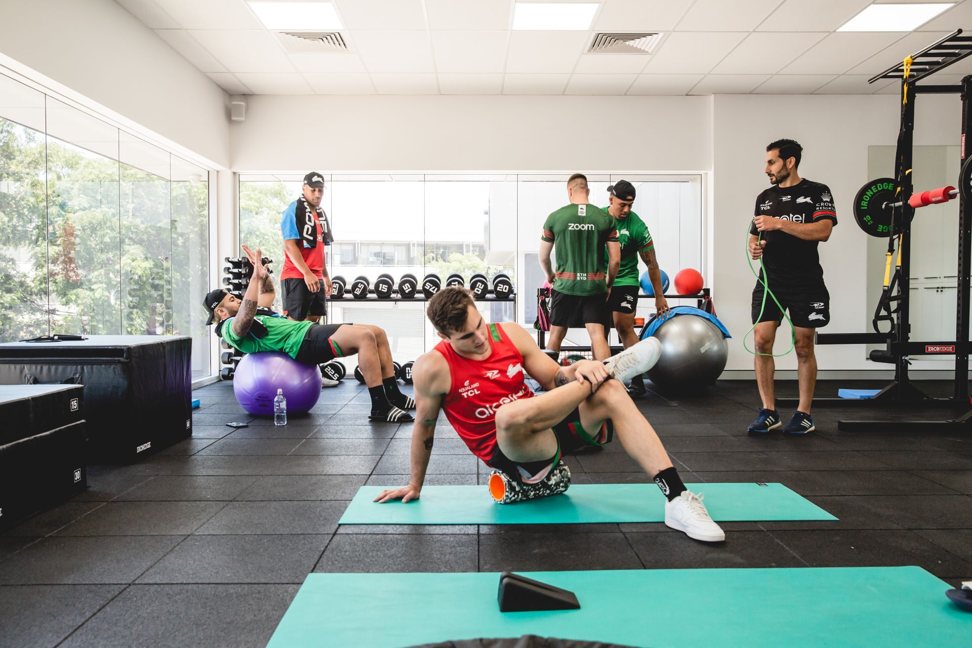
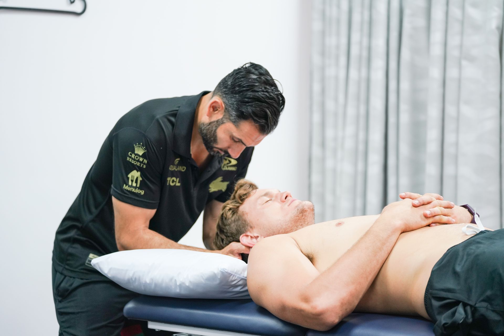
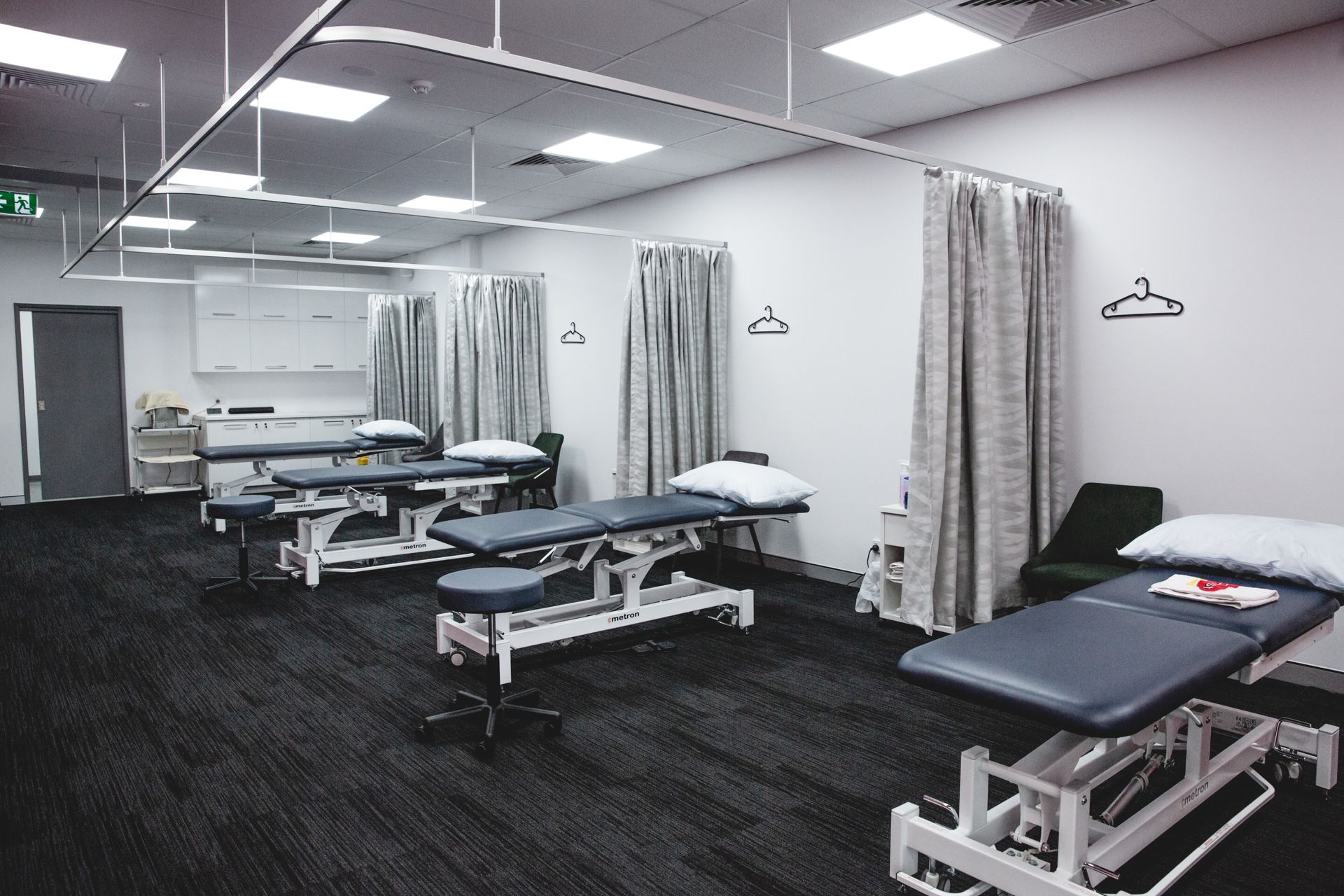
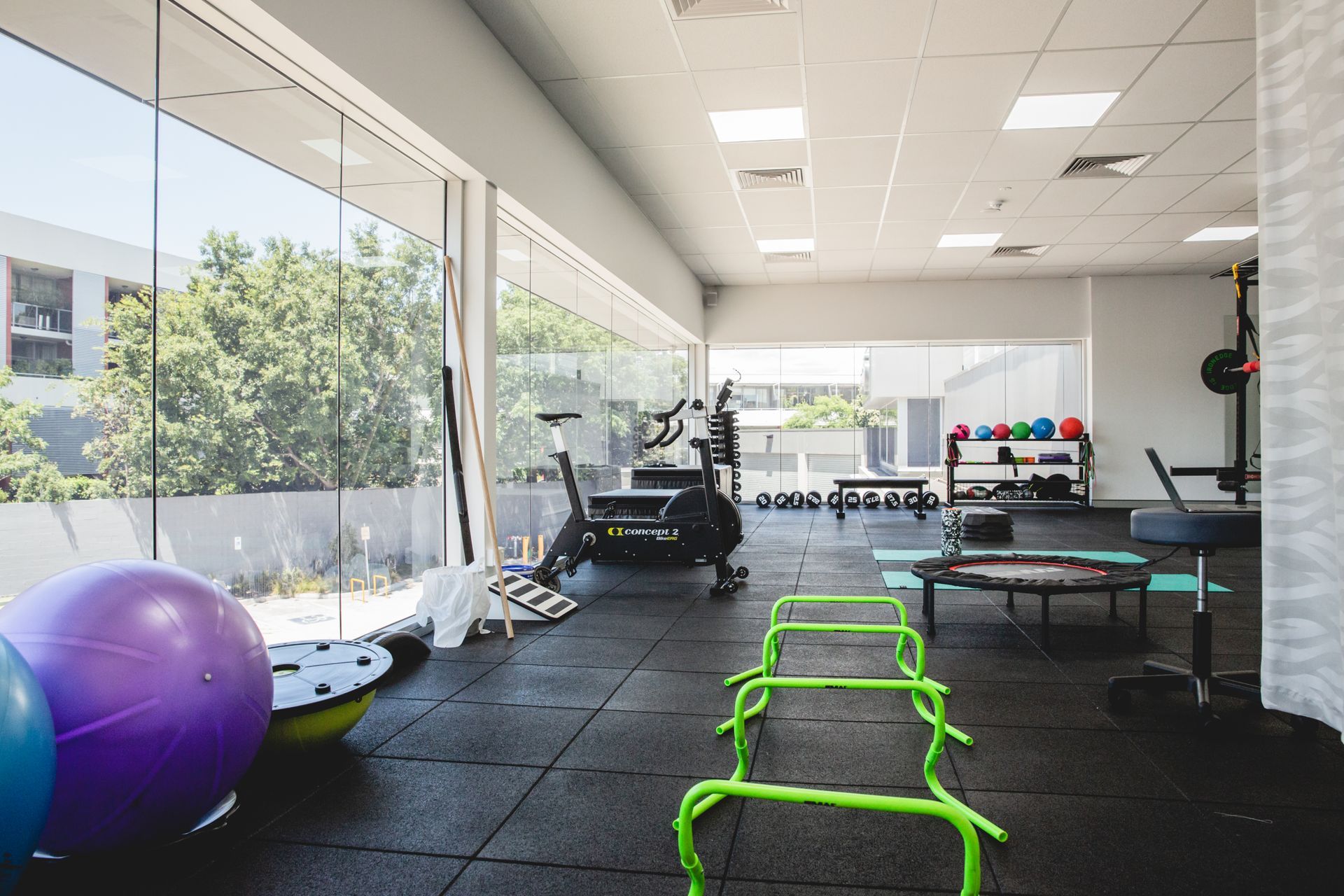
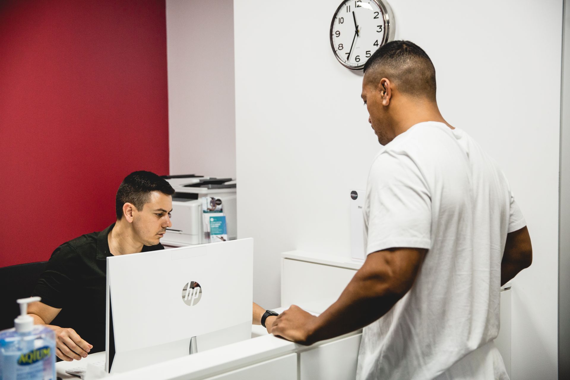
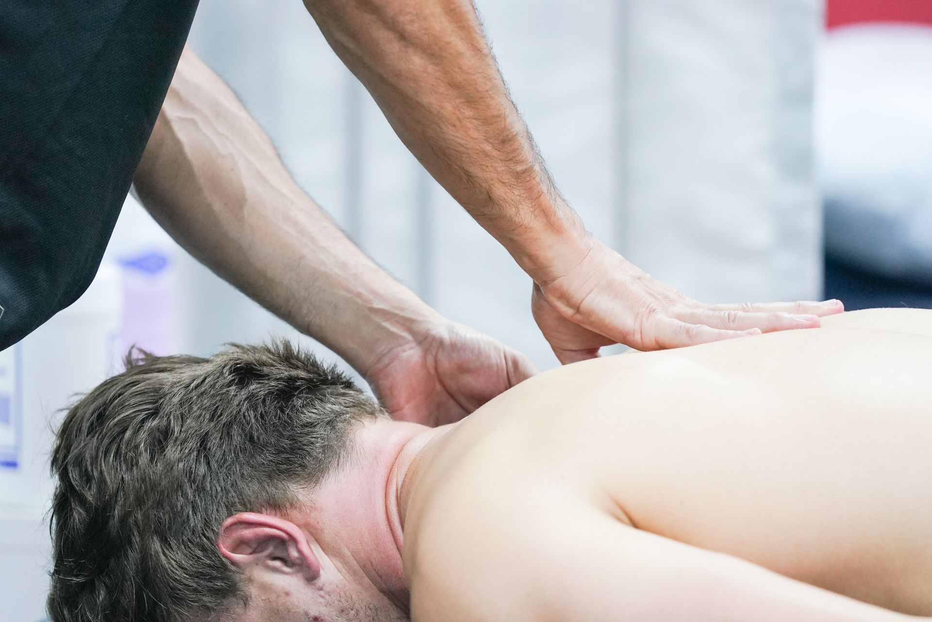
Gallery


View more